Fluorescence Spectroscopy
Fluorescence spectroscopy is an analytical technique that studies the light emitted by certain molecules after they absorb radiation. Unlike infrared spectroscopy, which measures the absorption of infrared light, or Raman spectroscopy, which measures scattered light, fluorescence spectroscopy measures light that is first absorbed and then re-emitted by a molecule. Because fluorescence can be extremely sensitive, it has become one of the most powerful techniques for chemical analysis, biological imaging, medical diagnostics, environmental monitoring, and pharmaceutical research.
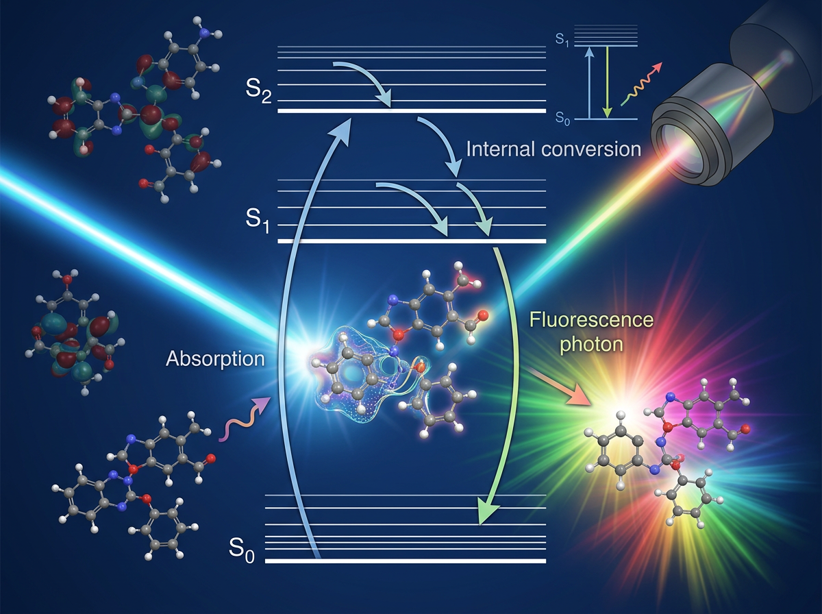
The process begins when a molecule absorbs a photon, usually from an ultraviolet (UV) or visible light source. The energy of the incoming photon promotes an electron from the ground electronic state (S0) to an excited electronic state (S1, S2, or higher). This absorption event occurs almost instantaneously, typically within femtoseconds.
However, molecules do not usually remain in these highly excited states. Almost immediately after excitation, they lose a portion of their excess energy through non-radiative processes such as molecular vibrations and collisions with surrounding molecules. As a result, the electron rapidly relaxes to the lowest vibrational level of the first excited electronic state (S1). From this state, the electron can return to the ground state by emitting a photon. This emitted light is called fluorescence.
Since some energy has already been lost through vibrational relaxation before emission occurs, the emitted photon possesses less energy than the absorbed photon. Consequently, fluorescence emission appears at a longer wavelength and lower energy than the excitation light. This difference between the excitation wavelength and emission wavelength is known as the Stokes shift and is one of the defining characteristics of fluorescence spectroscopy.
A useful way to visualize fluorescence is through the Jablonski diagram. In this diagram, vertical arrows represent absorption of light, where an electron is promoted to a higher electronic state. Curved downward arrows represent rapid vibrational relaxation that occurs without the emission of light. Finally, a downward straight arrow from the excited state back to the ground state represents fluorescence emission. The emitted photon carries information about the molecule's electronic structure and local environment, making fluorescence a highly informative analytical signal.
Not all molecules fluoresce. For fluorescence to occur efficiently, the molecule typically must contain electronic structures capable of absorbing UV or visible light. Aromatic compounds, conjugated systems, dyes, fluorescent proteins, and many biological molecules are common fluorophores because their delocalized electrons can be excited relatively easily. Molecules lacking suitable electronic transitions often do not exhibit measurable fluorescence.
One of the most important concepts in fluorescence spectroscopy is fluorescence intensity. The intensity of emitted light depends on several factors, including the concentration of the fluorophore, the excitation intensity, and the efficiency with which absorbed energy is converted into fluorescence. This efficiency is known as the quantum yield. A molecule with a high quantum yield emits a large fraction of absorbed energy as fluorescence, producing a bright signal. Molecules with low quantum yields lose most of their energy through non-radiative processes and therefore fluoresce weakly.
Because fluorescence emission is often measured against a very low background signal, fluorescence spectroscopy can achieve remarkable sensitivity. While many spectroscopic techniques require relatively large amounts of material, fluorescence measurements can often detect trace concentrations and, in specialized experiments, even individual molecules. This extraordinary sensitivity has made fluorescence one of the most valuable tools in modern analytical science.
The fluorescence spectrum itself provides useful molecular information. The position of the emission peak depends on the electronic structure of the molecule, while changes in intensity or peak shape can reveal information about the surrounding environment. Factors such as solvent polarity, pH, temperature, ionic strength, and interactions with other molecules can influence fluorescence behavior. For this reason, fluorescent probes are widely used as molecular sensors in biological and chemical systems.
An important competing process is phosphorescence, which is often confused with fluorescence. In fluorescence, the emitted light typically appears within nanoseconds after excitation and originates from transitions between states of the same spin multiplicity. In phosphorescence, the excited electron undergoes a spin change and enters a triplet state before emission occurs. Because this transition is less favorable, phosphorescence may persist for milliseconds, seconds, or even longer after the excitation source has been removed. Common "glow-in-the-dark" materials exhibit phosphorescence rather than fluorescence.
Modern fluorescence spectrometers are relatively straightforward in design. A light source, often a xenon lamp, LED, or laser, provides excitation radiation. Optical filters or monochromators select the desired excitation wavelength and direct it onto the sample. The emitted fluorescence is collected at an angle, usually 90 degrees from the excitation beam, minimizing interference from transmitted excitation light. A detector then records the emitted photons and generates the fluorescence spectrum.
Today, fluorescence spectroscopy is used across numerous scientific disciplines. In biology and medicine, fluorescent dyes and proteins allow researchers to visualize cells, tissues, and biomolecules with extraordinary sensitivity. In analytical chemistry, fluorescence measurements are used to quantify trace compounds and monitor chemical reactions. In environmental science, fluorescent methods can detect pollutants at extremely low concentrations. In pharmaceutical research, fluorescence assays are routinely employed for drug discovery and molecular interaction studies.
At its core, fluorescence spectroscopy is a remarkably elegant process. A molecule absorbs a photon, briefly stores the energy in an excited electronic state, and then releases part of that energy as light. By carefully measuring this emitted light, scientists gain valuable insights into molecular structure, concentration, dynamics, and interactions, making fluorescence spectroscopy one of the most versatile and sensitive techniques in modern science.